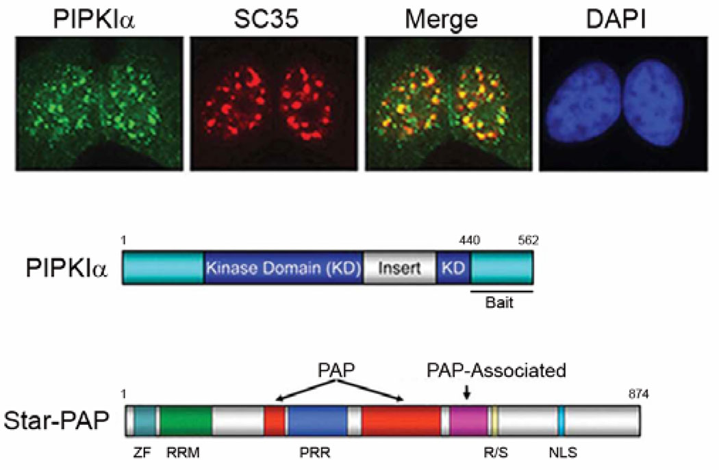
Fig. 2.
Endogenous PIPKIα localizes at nuclear speckles (top panel) evidenced by the co-localization of PIPKIα (green) with the SR protein SC35 (red). Merge shows co-localization. 4′,6-diamidino-2-phenylindole (blue). Amino acids 440–562 of PIPKIα was used as bait in a yeast two-hybrid screen to identify PIPKIα interacting proteins that function together in nuclear events (middle panel). Star-PAP was one of numerous nuclear localized proteins isolate din the screen and has been shown to function with PIPKIα in the 3’ end formation of select mRNAs (bottom panel) (ref. Mellman et. al., 2008).

