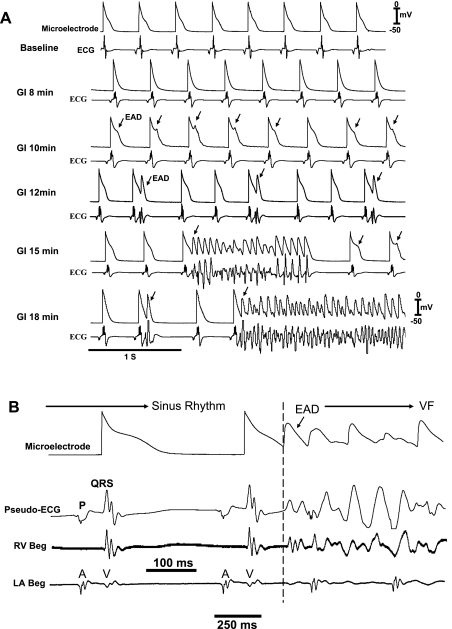
Fig. 2.
Time course of emergence of epicardial early afterdepolarizations (EADs), triggered activity, and VF in an aged heart exposed to GI. A: simultaneous microelectrode (top) and pseudo-ECG (bottom) recordings at baseline and at increasing time (18 min) after GI. EADs emerge 10 min after GI after APD shortening (8 min post-GI); EAD-mediated single triggered APs arise after 12 min causing premature ventricular depolarization as seen on the pseudo-ECG, which then evolve into short runs of triggered activity causing VT (15 min), which then degenerates to VF (18 min post-GI). B: epicardial EAD emerges when the ECG and the right ventricular (RV Beg) and left atrial (LA Beg) electrograms manifest isoelectric interval, indicating absence of electrical activity elsewhere in the heart.

