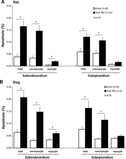
Fig. 1.
A: apoptosis in rat model of transverse aortic constriction (TAC). This figure compares the apoptosis (%) in left ventricular hypertrophy (LVH) after 4 wk of TAC (filled bars) vs. sham-operated rats (open bars) in the subendocardium (left) and subepicardium (right) for myocytes and for nonmyocytes. Apoptosis increased significantly more in LVH for both nonmyocytes and myocytes subendocardially (left) and for nonmyocytes subepicardially (right). B: dog model of TAC. The data resembled those from the rat model. Apoptosis is compared in 4 wk TAC with sham (*P < 0.05).

