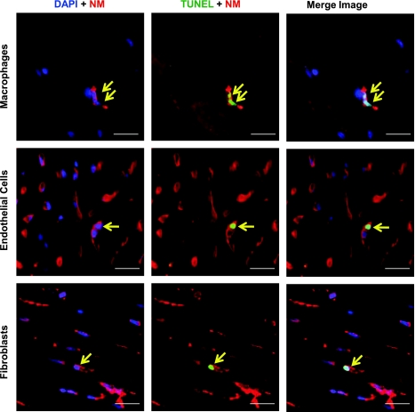
Fig. 3.
Characterization of cell types for apoptotic nonmyocytes in rat LVH. Representative photographs of apoptotic nonmyocytes (arrows) by double staining with TUNEL and specific antibodies (CD68 for macrophages, isolectin GS-IB4 for endothelial cells, and HSP47 for fibroblasts) using ×60 magnification. Left: nonmyocyte with DAPI. Middle: TUNEL-positive nonmyocyte. Right: the merged image of an apoptotic nonmyocyte. Green: TUNEL; red: nonmyocytes (NM); blue: DAPI. Scale bar: 20 μm.

