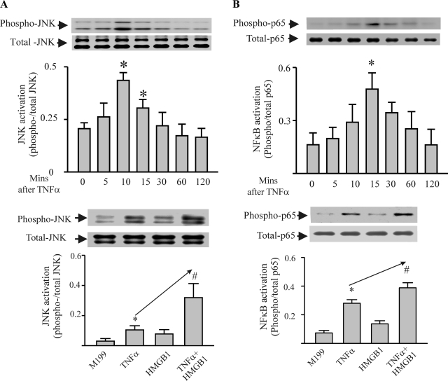
Fig. 4.
HMGB1 significantly potentiates TNF-α-induced activation of JNK, but not NF-κB in cardiac myocytes. A, top: time course of TNF-α-induced activation of JNK (JNK phosphorylation). TNF-α (40 ng/ml) was added to cardiomyocytes, and, at the times indicated, the myocytes were harvested for assessment of the phosphorylation status of JNK by Western blot. n = 3. *P < 0.05 compared with time 0. Bottom: cardiac myocytes were treated with M199 or one of TNF-α (40 ng/ml), HMGB1 (1 μg/ml), or TNF-α/HMGB1 cocktail for 10 min. The cells were harvested for assessment of the phosphorylation status of JNK by Western blot. n = 3. *P < 0.05 compared with M199. #P < 0.05 compared with TNF-α. B, top: time course of TNF-α-induced activation of NF-κB (p65 phosphorylation). Cardiac myocytes were treated with TNF-α as above and harvested for assessment of the phosphorylation status of p65 by Western blot at the times indicated. n = 3. *P < 0.05 compared with time 0. Bottom: cardiac myocytes were treated with M199 or one of TNF-α (40 ng/ml), HMGB1 (1 μg/ml), or TNF-α/HMGB1 cocktail for 15 min. The cells were harvested for assessment of the phosphorylation status of p65 by Western blot. n = 3. *P < 0.05 compared with M199. #P < 0.05 compared with TNF-α. Values are means ± SE.

