Figure 3.
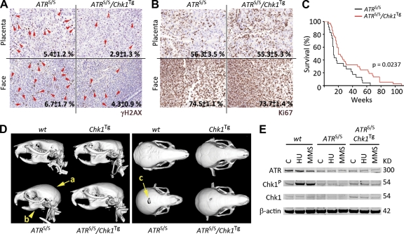
Alleviation of the ATR-Seckel syndrome by Chk1Tg. (A and B) Representative images of γH2AX (A) and Ki67 (B) on the placenta and face from ATRS/S and ATRS/S/Chk1Tg littermate embryos. Red arrows indicate cells showing a pan-nuclear γH2AX staining. Numbers indicate the mean percentage and SD of positive cells in each case (n = 3). (C) Kaplan-Meyer curves of ATRS/S (n = 23) and ATRS/S/Chk1Tg (n = 36) mice. The p-value was calculated with the Mantel-Cox log-rank test. (D) Computerized Tomography-mediated reconstruction of the heads from WT, Chk1Tg, ATRS/S, and ATRS/S/Chk1Tg mice. Yellow arrows indicate features that show evident rescue such as the shape of the crania (a), the micrognathia (b), and the deficient closure of the fontanelle (c). Data are representative of four independent analyses. (E) ATR, Chk1-P, and Chk1 protein levels in WT, ATRS/S, and ATRS/S/Chk1Tg MEF treated with 0.5 mM HU for 3 h or 10 mM methyl methanesulfonate (MMS) for 4 h. β-Actin was used as a loading control. Data are representative of two independent analyses.

