Figure 10.
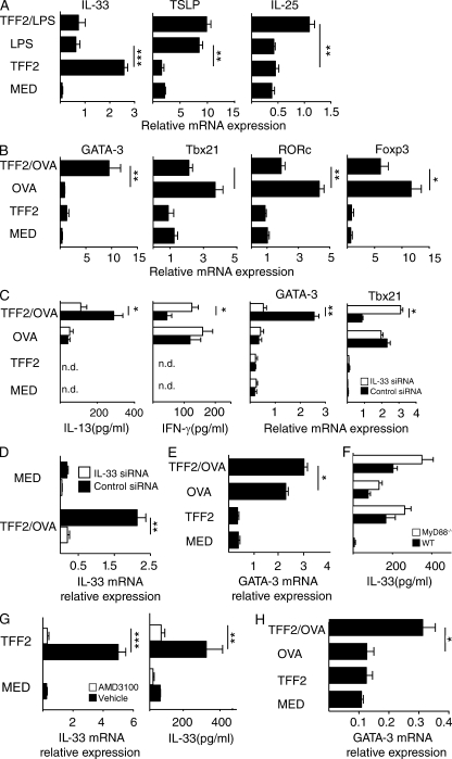
TFF2-treated antigen-presenting cells selectively drive TH2 differentiation through an IL-33–dependent mechanism. (A) BMDMs from WT mice were evaluated for mRNA expression levels of IL33, IL25, and TSLP after exposure to media (MED), 100 ng/ml LPS, 40 ng/ml rhTFF2 (TFF2), or LPS and TFF2 for 48 h. Mean ± SE of quadruplicate wells is shown. (B) Quantification of mRNA transcripts for GATA3, TBX21, RORC, and FOXP3 in co-cultures of OTII CD4+ T cells and BMDMs that were left untreated (MED), treated with 40 ng/ml rhTFF2, pulsed with 50 µg/ml OVA, or pretreated with rTFF2 and pulsed with 50 µg/ml OVA (TFF2 + OVA). Cells were analyzed at 96 h. Data show mean ± SE of triplicate wells. (C) BMDMs were transfected with siRNA specific for IL-33 (IL-33 siRNA) or scrambled control (control) 48 h before the co-culture experiment described in B. IL-13 and IFN-γ protein levels were determined by ELISA, and mRNA levels of GATA3 and TBX21 were determined by quantitative RT-PCR. Mean ± SE of triplicate wells is shown. (D) BMDMs transfected with scrambled control or IL-33 siRNA were tested for IL-33 induction 96 h after treatment with OVA and 40 ng/ml rTFF2. Mean ± SE of triplicate wells is shown. (E) GM-CSF differentiated BMDCs from naive WT C57BL/6 mice were stimulated as in B. Error bars indicate SE. (F) IL-33 protein levels in supernatants of WT versus MyD88−/− BMDMs that were subjected to the co-culture conditions described in B. The experiment was performed two times. Data show mean ± SE of triplicate wells. (G) Il33 mRNA expression and IL-33 protein levels from WT BMDMs that were exposed to 1 µg/ml AMD3100 (CXCR4 antagonist) 24 h before exposure to stimulation with 40 ng/ml TFF2 or media for 48 h. Data show mean ± SE of triplicate wells. (H) GATA3 expression levels from co-cultures of WT naive CD4+ OTII cells and TI/ST2−/− BMDMs that were treated with 40 ng/ml rTFF2 or left untreated before pulse with OVA (endotoxin <0.2 ng/ml free) and co-cultured with OTII cells for 96 h. Data show mean ± SE from triplicate wells. Data are representative of two to four independent experiments (*, P < 0.05; **, P < 0.01; and ***, P < 0.001).

