Figure 8.
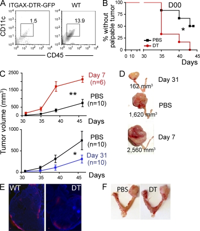
Distinct populations of DCs promote immunosurveillance and the escape phase of tumor development. (A) FACS analysis of dissociated ovaries from mice reconstituted with ITGAX-DTR-GFP or WT BM, 24 h after the i.p. administration of 6 ng/kg of diphtheria toxin. (B) Proportion of p53/K-ras mice reconstituted with BM from ITGAX-DTR-GFP mice without palpable tumors at the indicated times after administration of 6 ng/g of diphtheria toxin (DT) or PBS, 1 d before adenoviral injection (n = 6mice/group). (C) p53/K-ras mice were reconstituted with BM from ITGAX-DTR-GFP mice, and DCs were depleted with one dose of 6 ng/kg of diphtheria toxin (DT) at days 7 (n = 6 mice/group, top), or 31 (n = 10/group, bottom; two pooled independent experiments) after intrabursal adenovirus-Cre. PBS (n = 10/group; two pooled independent experiments) was administered to control mice. (D) Representative size of advanced ovarian tumors in mice depleted of DCs at early versus advanced stages, compared with the absence of DC depletion (PBS). Error bars, SEM. *, P < 0.05; **, P < 0.01. (E) ITGAX-DTR (DT) or WT mice (n = 3/group) were inoculated intrabursally with 2.5 × 10⁷ plaque-forming units of adenovirus expressing Red-Cherry, and red fluorescence was detected 4 d later. ITGAX-DTR mice received diphtheria toxin (6 ng/g body weight) 24 h before surgery. Brightness, contrast and color balance were uniformly adjusted in whole individual images. Bars, 100 µm. (F) Day 50 tumor growth in p53/Kras mice challenged with adenovirus-Cre and receiving i.p. PBS or diphtheria toxin (DT) 7 d later. Shown are representatives of four mice/group.

