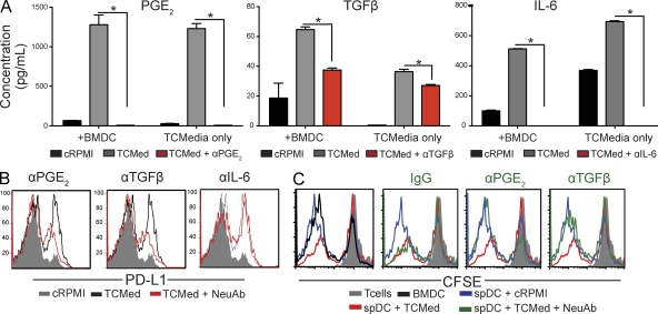
Figure 9.
Tumor cell–derived PGE2 and TGF-β1 promote the immunosuppressive activity of DCs. (A) Quantification of PGE2, mature TGF-β1, and IL-6 in media conditioned by tumor cells from advanced (∼60 d) ovarian cancer specimens, in the presence or the absence of specific neutralizing antibodies. +BMDCs, BMDCs were incubated for 2 d in tumor-conditioned media before cytokine quantification; cRPMI, RPMI control media; TCmedia, tumor-conditioned media plus an irrelevant IgG; TCmedia+NeuAb, tumor-conditioned media plus specific neutralizing antibodies. Error bars, SEM. *, P < 0.05. (B) PD-L1 expression in splenic CD45+CD11c+MHC-II+ DCs sorted from mice carrying early (day 7) tumor lesions, cultured with control versus tumor-conditioned media, in the presence or the absence of specific neutralizing antibodies. (C) Splenic DCs (spDCs) sorted from early tumor-bearing mice were cultured for 48 h in RPMI or tumor-conditioned media (TCMed), in the presence of neutralizing anti-PGE2 or anti–TGF-β1 antibodies (NeuAb), or an irrelevant IgG. These cultured DCs were then incubated with tumor-pulsed BMDCs plus CFSE-labeled tumor-reactive T cells, obtained as in Fig. 6 A (1:1:1 ratio). Unpulsed BMDCs were added to control wells. The panel shows representative histograms of the proliferation of tumor-reactive T cells under different conditions. Representatives of two independent experiments are shown.

