
Fig 3.
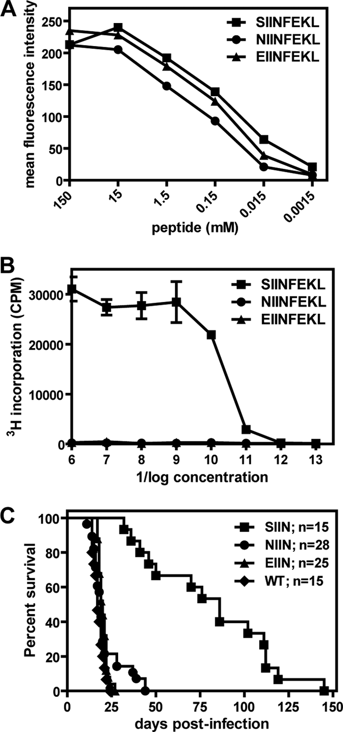
NIINFEKL peptide binds to MHC class I but fails to stimulate OTI T cells. (A) RMA-S cells were pulsed with various concentrations of the SIINFEKL, NIINFEKL, or EIINFEKL peptide and stained with the 25.D1.16 monoclonal antibody. H2-Kb cell surface expression was assessed by flow cytometry. The data are representative of results from two independent experiments. (B) OTI T cells were incubated with γ-irradiated splenocytes that had been pulsed with various concentrations of the SIINFEKL, NIINFEKL, or EIINFEKL peptide. After 24 h of coincubation, 3H-thymidine was added for an additional 12 h of incubation. 3H-thymidine incorporation was quantitated as a measure of OTI T cell proliferation. The samples were analyzed in triplicate, and these data are representative of the results of 2 independent experiments. (C) OTI-Rag mice were infected with BAC-derived wild-type γHV68 (WT) or OVA-expressing viruses containing the SIINFEKL (SIIN), NIINFEKL (NIIN), or EIINFEKL (EIIN) epitope. Insertion of the OVA expression cassette into each BAC recombinant virus, as well as the lack of gross mutations elsewhere in the viral genomes, was confirmed by Southern blot analyses (not shown). The mice were infected i.p. with either 4 × 10³ or 5 × 10³ PFU/mouse and were followed for lethality. These data represent the results of at least 2 independent experiments, and the total numbers of mice analyzed are indicated.