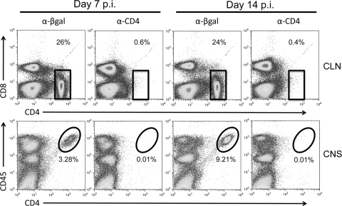
Fig 2.
α-CD4 MAb treatment prevents CD4 T cell accumulation in the CNS. JHMV-infected mice were treated with α-CD4 or control MAb at 4 and 6 days p.i. Pooled CLN (upper row) or brain cells (bottom row) (n ≥ 3/group) isolated at days 7 and 14 p.i. were stained for CD4, CD8, or CD45. Percentages of CD4+ T cells within the CLN (boxed cells) are shown in the upper right corner. Percentages of CD4+ T cells within the CNS (circled cells) are shown below the ellipse. Data are representative of results of three independent experiments.

