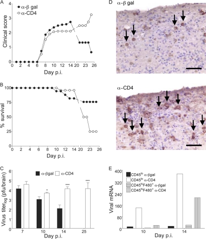
Fig 3.
Delayed depletion of CD4 T cells impairs viral control in the CNS. JHMV-infected mice were treated with α-CD4 or control MAb at 4 and 6 days p.i. and monitored for clinical symptoms (A), survival (B), and virus titers in brains by plaque assay (C). Titers are expressed as the means ± SEM. (D) Virus-infected cells in spinal cord white matter tracks of infected mice at 10 days p.i. Immunoperoxidase stain using α-nucleocapsid MAb (brown) with hematoxylin counterstain. Arrows indicate viral nucleocapsid Ag-positive cells with morphology consistent with oligodendroglia. Scale bar, 100 μm. (E) Relative transcript levels of viral nucleoprotein in purified CD45lo microglia and CD45hi F4/80+ monocyte-derived macrophages from pooled brains (n = 6) 10 and 14 days p.i. were assessed by real-time PCR. Transcript levels are relative to GAPDH. Statistically significant differences in overall clinical score up to day 14 p.i. between control and CD4 T cell-depleted mice were determined by Wilcoxon matched pairs test. **, P < 0.005. Statistically significant differences in viral titers between control and CD4 T cell-depleted mice were determined by unpaired t test. *, P < 0.5; ***, P < 0.001.

