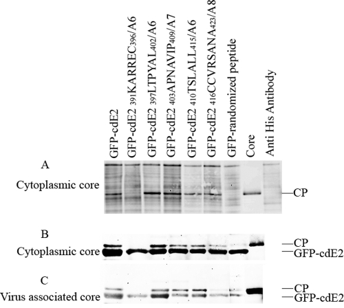
Fig 6.
Coimmunoprecipitation analysis of cdE2 fusion proteins with CP, core-like particles, and cytoplasmic cores. (A) Coimmunoprecipitation (co-IP) of CP and GFP-cdE2. The cytoplasmic cores purified from SINV-infected cells were mixed with GFP-cdE2 or mutant proteins, and the GFP-cdE2 and NC complexes were immunoprecipitated using anti-penta-His mouse monoclonal antibody directed against the N-terminal His tag of GFP. The proteins were detected by Western blot analysis using anti-SINV-specific rabbit polyclonal anti-CP antibody and infrared-labeled goat anti-rabbit secondary antibody. The negative control lane, labeled “core,” represents the core used for the experiment with no antibody, and the anti-His antibody lane represents the antibody used in the pulldown experiment with no cores. (B) His tag pulldown assay of cytoplasmic cores using GFP-cdE2 fusion proteins. The proteins were pulled down using Profound pulldown poly-His protein-protein interaction kit (Pierce) against the N-terminal His tag of GFP and were analyzed by Western blotting using SINV-specific rabbit polyclonal anti-CP and mouse monoclonal anti-penta-His primary antibodies and infrared-labeled goat anti-rabbit and goat anti-mouse secondary antibodies. (C) His tag pulldown assay of virus-associated cores purified from detergent-treated SINV using GFP-cdE2 fusion proteins; experiments were conducted the same way as for B.

