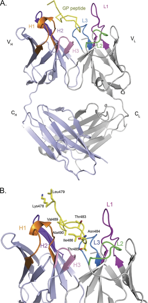
Fig 1.
Crystal structure of the 14G7 Fab-peptide complex. For clarity, only one of the two essentially identical molecules in the asymmetric unit is shown. (A) Overall structure of the 14G7 Fab in complex with its Ebola virus epitope. (B) Close-up side view of the Fab-peptide complex showing the spatial orientation of the peptide within the Fab pocket. The antibody CDRs L1, L2, L3, H1, H2, and H3 are colored magenta, green, blue, orange, purple, and pink, respectively, while the peptide is in yellow. All of the figures for the present study were generated by using MacPyMol (35).

