Fig 3.
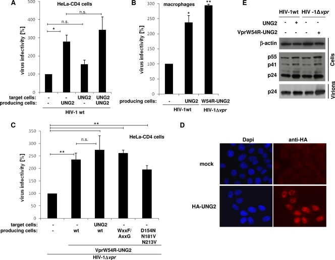
Impact of UNG2 overexpression on virus infectivity. (A and C) Wild-type or Δvpr single-round GFP reporter viruses carrying the HIV-1 HXBc2 envelope were produced in 293T cells overexpressing HA-tagged forms of UNG2, or wt or mutated VprW54R-UNG2 fusions, and were used to infect HeLa-CD4 cells overexpressing or not HA-UNG2. Viruses were normalized for CAp24 before infection. The percentages of GFP-positive infected cells were then measured by flow cytometry 60 h later. Viral infectivity was normalized to that of wt (in panel A) or Δvpr (in panel C) viruses produced in cells that did not overexpress UNG2 or VprW54R-UNG2. Values are the means of at least four independent experiments. Error bars represent one SD from the mean. Statistical significance was determined by using the Student t test (n.s., P > 0.05; *, P < 0.05; **, P < 0.01). (B) Wild-type or Δvpr GFP reporter viruses carrying the HIV-1 YU-2 envelope were produced in 293T cells overexpressing HA-tagged forms of either UNG2 or VprW54R-UNG2 and were used to infect primary macrophages derived from blood monocytes from three healthy donors. Viral infectivity was normalized to that of wt viruses produced in cells that did not overexpress UNG2 or VprW54R-UNG2. Values are the means of three independent experiments. Error bars represent one SD from the mean. Statistical significance was determined by using the Student t test (n.s., P > 0.05; *, P < 0.05, **; P < 0.01). (D) Overexpression of UNG2 in HeLa-CD4 target cells. Cells were transfected with the vector for the expression of the HA-tagged form of UNG2 (lower panels) and analyzed 24 h later by indirect immunofluorescence with anti-HA antibody (right panels). Nuclei were stained with DAPI (left panels). Cells were analyzed by epifluorescence microscopy, and images were acquired by using a charge-coupled device camera. (E) Virus maturation of wt and Δvpr HIV-1. 293T cells were cotransfected with wt or vpr-defective HIV-1-based vector in combination with plasmids for expression of HA-tagged forms of UNG2 or VprW54R-UNG2. Virions were collected from cell supernatants; proteins from cell and virion lysates were analyzed by Western blotting with anti-CAp24 and anti-β-actin as a control.

