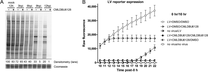
Fig 5.
(A) [35S]methionine labeling of cells to visualize active translation. Densitometry values are provided normalized to 12-h mock-infected cells (−). A representative slice of the Coomassie blue-stained gel is also shown. (B) Time course analysis of Venus expression in A549 cells subjected to various treatments. The legend indicates the treatments given at 0 h and 10 h. Reporter fluorescence was measured hourly for 12 h after the initial 10-h treatment (until 22 h). “LV” indicates high-MOI infection with the late Venus reporter virus. For example, LV+DMSO/CMLDBU6128 indicates an initial treatment of high-MOI LV reporter virus in the presence of DMSO, followed 10 h later by medium removal and replacement with CMLDBU6128-containing medium.

