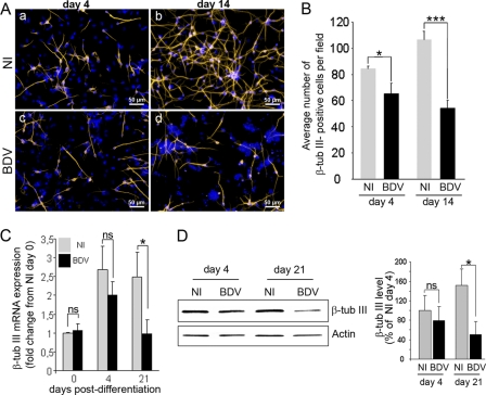
Fig 5.
BDV impairs neurogenesis. Fully infected HNPCs and their matched noninfected controls were induced to differentiate for 4 and 14 days by growth factor withdrawal. (A) Neuronal cells immunostained for β-tubulin III (orange) and DAPI (blue; nuclear staining). (B) Total number of β-tubulin III-positive cells in noninfected and BDV-infected cultures. Data represent mean values ± SEM from one experiment performed in triplicate. Similar results were obtained from 3 independent experiments. β-Tubulin III mRNA and protein were analyzed by quantitative real-time PCR (C) and Western blotting (D). Data represent mean values ± SEM from three independent experiments performed in duplicate (C) or two independent experiments performed in duplicate (D). Statistical analyses were performed by employing the Student's t test. ns, not significant; *, P < 0.1; ***, P < 0.005.

