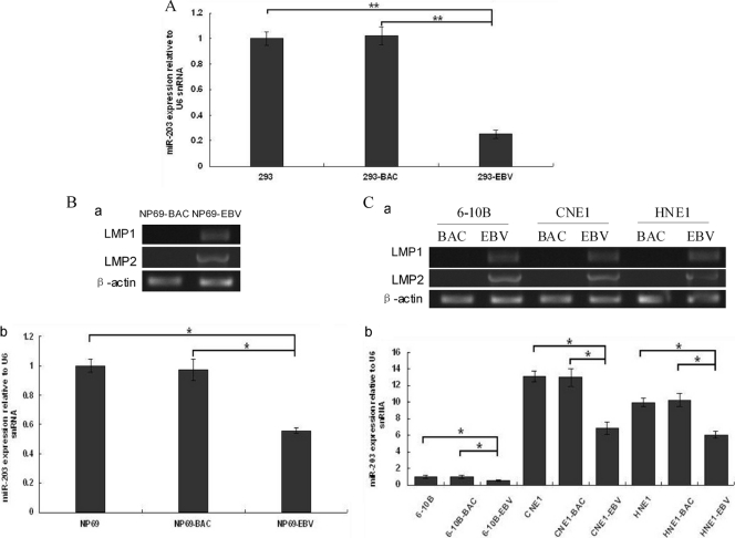
Fig 1.
miR-203 expression in EBV-infected epithelial cells. (A) In EBV stably infected 293-EBV cells, miR-203 was downregulated approximately 4-fold compared with the controls (293 and 293-BAC cells). 293-BAC cells were used as a negative control and were stably transfected with the BAC-based vector pM-BAC, which was established by eliminating the EBV genome from the Maxi-EBV plasmid. (B) Expression of latent genes and miR-203 in immortalized nasopharynx cells (NP69) during the first 7 days of infection using the transfer infection method with infectious EBV particles. The expression of LMP1 and LMP2 genes was determined using semiquantitative RT-PCR analysis (a). The expression of miR-203 was determined using real-time qRT-PCR analysis (b). NP69-BAC cells were used as a negative control and were transfected with the vector pM-BAC. (C) The expression of latent genes and miR-203 in nasopharyngeal carcinoma cells that were reinfected with EBV 10 days postinfection. Expression of LMP1 and LMP2 (a) and of miR-203 (b) are shown. The 6-10B-BAC, CNE1-BAC, and HNE1-BAC cell lines were used as controls and were transfected with the vector, pM-BAC. For panels B and C, the EBV-infected cells were GFP positive under green fluorescence microscopy. miR-203 expression was normalized to that of U6, and the relative levels are shown, with the value of the blank-cell control of each group standardized to 1. The data correspond to the mean values of three independent experiments. A P value of <0.05 (*) or <0.01 (**) was considered to be statistically significant or extremely significant, respectively.

