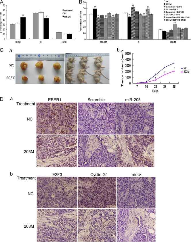
Fig 6.
Ectopic expression of miR-203 reversed the phenotype changes of EBV infection. (A) Cell cycle distribution after ectopic expression of miR-203 in 293-EBV cells. Two days after transfection with miR-203 mimics (203 M) and scramble (NC), cells were harvested, fixed with ethanol, and subjected to the measurement by flow cytometry. (B) The overexpression of E2F3 and/or CCNG1 together with miR-203 overcame the effect of miR-203 on the cell cycle distribution. The 293-EBV cells were cotransfected with expression vectors, scramble and the vector pCMV6-XL (for NC), or miR-203 mimics (203 M) (20 nM) and pCMV6-XL (for 203 M), or 203 M/scramble and E2F3/CCNG1 (a mixture of E2F3 and CCNG1) (for the other six groups). Each expression vector contained the entire coding sequence of the gene minus the 3′ UTR. For panels A and B), the values are shown as the mean of three independent experiments (*, P < 0.05 compared with the NC values). (C) The effect of miR-203 on the growth of tumors formed by the 293-EBV cells. Three million cells were injected into mice; three of four mice from each group formed tumors at the site of inoculation. A tumor was first detected at day 7 postinjection. During the intratumoral injection of the 203 M or NC, the xenograft tumor volume was examined. The graph in panel a shows tumor formation in the nude mice. The graph in panel b shows the curve of tumor growth during the treatment with 203 M or NC (*, P < 0.05). The average tumor volume in the 203 M group (n = 3) was statistically significant from the volume in the mice treated with NC. (D) The expression of miR-203 and the two target genes in the tumor tissues at the end of the 203 M intratumoral injection treatment. Serial sectioned specimens were used. The expression of EBER1 and miR-203 (a) was determined by ISH. Scramble was used as a negative control, which was a probe attached to a scrambled sequence. The expression of E2F3 and CCNG1 (b) was determined by immunohistochemical analysis. Mock, no primary antibody.

