Fig 1.
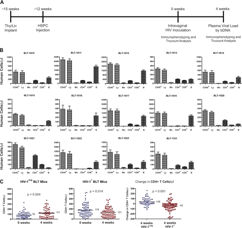
Experimental design, HIV inoculation, and loss of CD4+ T cells in HIV-infected BLT mice. (A) Fourteen cohorts of BLT mice (Table 1) were inoculated intravaginally with HIV JR-CSF at 0 weeks (12 weeks after HSPC injection). Peripheral blood of BLT mice was evaluated for human cell reconstitution by Trucount analysis 0 weeks and 4 weeks after inoculation. bDNA, branched DNA assay. (B) Total numbers of peripheral blood leukocytes (CD45+), lymphocytes (Ly), monocytes (Mo), CD4+ T cells (CD4+), CD8+ T cells (CD8+), and B cells (B) were determined for each cohort by Trucount analysis at the time of inoculation. (C) Significant differences were observed in the number of CD4+ T cells and the change in CD4+ T-cell number between HIV-negative (n = 58) and HIV-positive (n = 79) mice at the time of inoculation and 4 weeks after inoculation. The Mann-Whitney U test was used to test the differences between the means.

