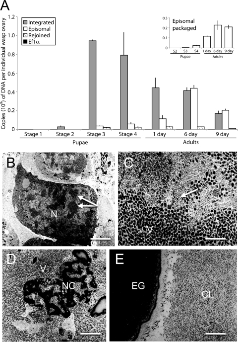
Fig 1.
Timing of proviral segment B amplification, excision/circularization of episomal segment B, and MdBV virion formation in M. demolitor. (A) Mean copy number ± standard error (SE) of qPCR products corresponding to integrated segment B, episomal segment B, the empty segment B locus, and EF1α. The total number of copies of each DNA in an individual wasp ovary is presented along the y axis, while the stages of wasp development are presented along the x axis. The inset shows mean copy number ± SE of episomal segment B at each wasp stage that is packaged into virions. The data for each wasp stage derives from two or three independently collected ovary samples. (B to E) TEM micrographs of MdBV nucleocapsids and virions during particular phases of replication. (B) Low-magnification image of calyx cells from a stage 3 pupa. Note the enlarged nucleus (N) for the calyx cell in the center of the image. Arrows indicate regions in the nucleus where MdBV particles are being assembled in proximity to virogenic stroma. Bar = 1.5 μm (C) High-magnification image of a calyx cell nucleus from a stage 3 pupa. The arrow indicates a region where both empty viral envelopes and envelopes containing a capsid are visible, while below this region are an abundance of mature virions (V). Bar = 100 nm. (D) High-magnification image of a lysed calyx cell from a stage 4 pupa. The nuclear membrane of the cell has deteriorated, resulting in release of mature virions (V) and dense nuclear chromatin (NC). Bar = 500 nm. (E) Low-magnification image from a day 1 adult showing densely packed MdBV virions in the calyx lumen (CL) that is adjacent to an M. demolitor egg (EG). Bar = 1 μm.

