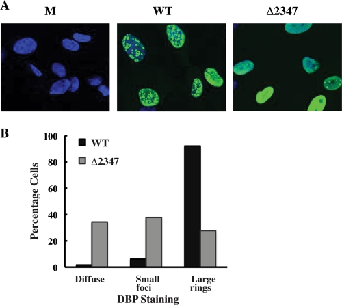
Fig 2.
Formation of viral replication centers in AdEasyE1- and AdEasyE1Δ2347 mutant-infected HFFs. (A) The E2 DBP was examined by immunofluorescence (as described in Materials and Methods) 25 h after infection of HFFs with 50 PFU/cell AdEasy E1 (WT), the AdEasyE1Δ2347 mutant (Δ2347), or mock-infected cells (M). Nuclei were stained with DAPI (4′,6-diamidino-2-phenylindole) (blue). (B) The appearance of DBP only as diffuse nuclear staining (diffuse), in small dot-like foci with or without diffuse DBP (small foci), or in enlarged ring-like structures (large rings) was counted in ≥100 cells infected by AdEasyE1 or the AdEasyE1Δ2347 mutant. The percentage of the total number of infected cells containing each form of DBP are shown.

