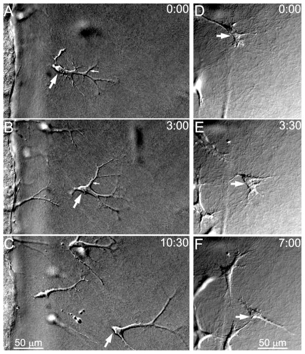
Figure 3.
Time-lapse DIC images of cell migration into bovine collagen outer matrices during culture in PDGF (A–C) or 10% FBS (D–F). A–C. PDGF induced repeated extension and retraction of long dendritic processes as cells migrated. Cells appeared to glide through the ECM without producing large deformations (See Supp Video 1). D–F. In 10% FBS, cells had thicker processes and were less elongated, and much stronger cell-matrix interactions were observed (See Supp Video 2). Large arrow points to same cell in each frame. Times in upper right corner show hours:minutes after starting time lapse imaging.

