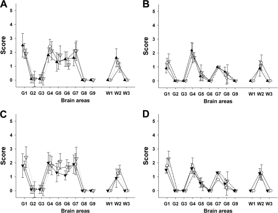
Fig 1.
Lesion profiles in tg338 mice inoculated with brain material and blood fractions from sheep. Shown are lesion profiles (showing vacuolar changes) in tg338 mice inoculated by the intracerebral route with (A) 10% obex homogenate prepared from clinically affected VRQ/VRQ sheep that were naturally exposed to Langlade classical scrapie agent (▴; blood donor 2 in Table 1) or that were transfused with blood collected from donor 2 at 90 days (○) and 360 days (▿) of age; (B) 10% obex homogenate from clinically affected VRQ/VRQ sheep that were orally challenged with PG127 classical scrapie agent (△; blood donor 5 in Table 2) or that were transfused with blood collected from donor 4 at 90 days (○) and 190 days (▿) postinoculation; (C) white blood cell homogenates (white symbols) and PBMC (black symbols) prepared from VRQ/VRQ sheep that were naturally exposed to Langlade classical scrapie agent (blood donor 2 in Table 3) collected at 210 days (▿) and 360 days (○) of age; and (D) white blood cell homogenates (white symbols) and PBMC (black symbols) prepared from VRQ/VRQ sheep that were orally exposed to PG127 classical scrapie agent (blood donor 5 in Table 4) and collected 130 days (▿) and 180 days (○) postinoculation.

