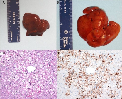
Fig 7.
Gross, histologic, and immunohistochemical findings in the liver of RVFV-infected marmosets. (A) Gross image of the liver of an uninfected control marmoset. (B) Gross image of the liver of an RVFV-infected marmoset exposed i.v. on day 2 p.i. (C) Hematoxylin and eosin stain. Hepatocellular degeneration and necrosis in the liver on day 4 p.i. of a marmoset exposed s.c. are shown. (D) Immunohistochemistry demonstrating the amount of RVFV antigen (brown staining) on day 4 p.i. in the liver (primarily degenerate/necrotic hepatocytes) of a marmoset exposed s.c.

