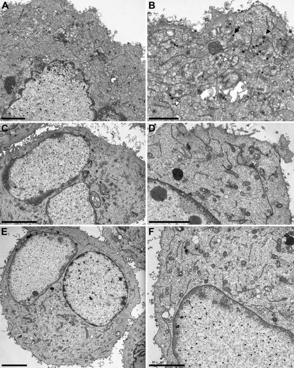
Fig 7.
Ultrastructural analysis of Lap2ß-Chimera. RK13-UL34-LapCT50 (A and B), RK13-UL34-LapCT100 (C and D), and RK13-UL34-LapNT (E and F) were infected with PrV-ΔUL34 at an MOI of 1 and processed for electron microscopy 14 h after infection. On the left overviews of infected cells are shown, and higher magnifications are given on the right. Bars correspond to 2 (A), 1.4 (B), 5 (C), 3 (D), 5 (E), and 3 μm (F). In panel B, the primary enveloped virion is marked by an asterisk, an intracytoplasmic nucleocapsid by an arrow, an intracytoplasmic nucleocapsid undergoing secondary envelopment by a closed triangle, and an enveloped intracytoplasmic virion in a vesicle by an open triangle. No extranuclear virus particles are present in panels C to F, whereas intranuclear DNA-filled capsids are clearly visible.

