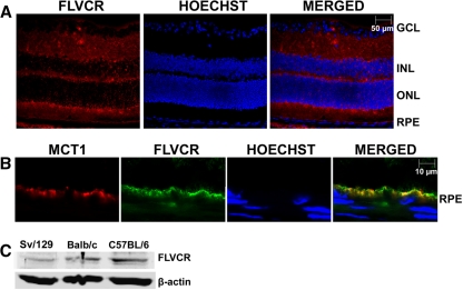
Figure 2.
Expression of FLVCR protein in mouse retina and its polarized localization in RPE. (A) Immunohistochemical localization of FLVCR in 3-month-old mouse (BALB/c) retina. Hoechst was used as a nuclear stain. BALB/c mice were used because of the absence of pigment in the RPE (the RPE pigment autofluoresces, thus making the interpretation of immunofluorescence data difficult). GCL, ganglion cell layer; INL, inner nuclear layer; ONL, outer nuclear layer. (B) Co-localization of MCT1 (an apical membrane marker in RPE) and FLVCR in RPE. (C) Western blot analysis of FLVCR protein in lysates prepared from retinas of Sv/129, BALB/c, and C57BL/6 mice.

