Fig 3.
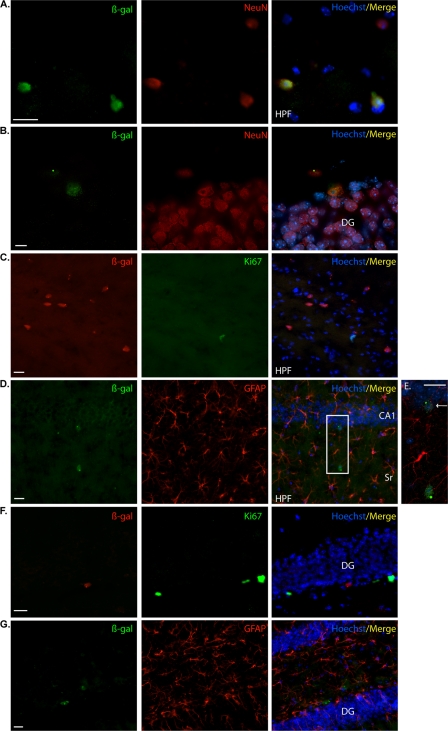
NR2E1-lacZ targeted at the Hprt locus demonstrated an unexpected absence of expression in adult proliferative cells of the dentate-gyrus-subgranular layer. (A) In BAC NR2E1-lacZ mice, immunofluorescence using an anti-β-Gal antibody (green) revealed positive cells in the hippocampal formation (HPF) that colocalized with NeuN (red), suggesting expression in mature neurons. (B) Colocalization revealed few β-Gal-immunoreactive cells (green) in the subgranular layer of the dentate gyrus (DG) that colocalized with NeuN (red), suggesting expression in mature neurons. (C) β-Gal-immunopositive cells (red) in the HPF did not colocalize with Ki67 (green). (D) β-Gal-immunopositive cells (green) in the HPF did not colocalize with GFAP (red). The boxed region in panel D is shown in panel E. (E) Higher magnification of the colocalization between β-Gal (green) and GFAP (red) revealed β-Gal-positive cells in cornu ammonis 1 (CA1) of the hippocampus (arrow). These cells did not colocalize with GFAP. (F) β-Gal-immunopositive cells (red) in the DG did not colocalize with Ki67 (green). (G) β-Gal-immunopositive cells (green) in the DG did not colocalize with GFAP (red). Sr, stratum radiatum. (Cryosection scale bars, 20 μm.)

