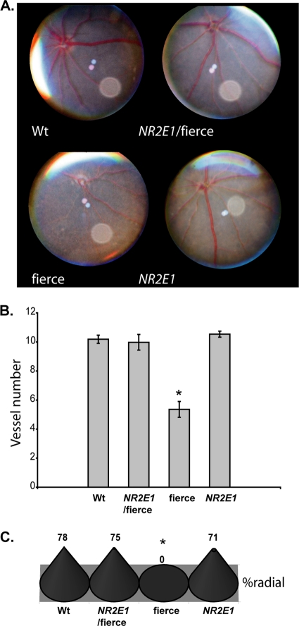
Fig 9.
NR2E1/fierce mice were corrected for retinal blood vessel defects. (A) Eye fundus photos showed normal blood vessel organization in the Wt, NR2E1/fierce, and NR2E1 retinal surface. The expected blood vessel abnormalities were seen for fierce mice. (B) No significant difference was found in the blood vessel numbers of Wt, NR2E1/fierce, and NR2E1 mice. The blood vessel number was significantly reduced in fierce eyes compared to the other mice (∗, P = 0.001). Error bars represent standard errors of the means. (C) No significant differences were found between Wt, NR2E1/fierce, and NR2E1 mice for asymmetry; only fierce mice showed asymmetry of the blood vessels (∗, P < 0.001). The Kruskal-Wallis H test was performed on 6 to 9 mice for all genotypes (B and C).

