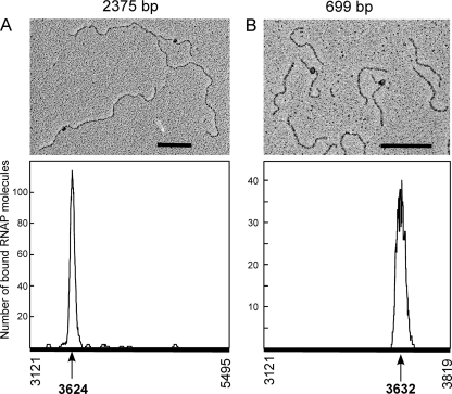
Fig 4.
Electron micrographs of RNAP-DNA complexes. The E. coli RNAP (120 nM) was incubated with the 2375-bp fragment (5 nM) (coordinates 3121 to 5495) (A) or with the 699-bp fragment (20 nM) (coordinates 3121 to 3819) (B). After 15 min at 37°C, complexes were fixed with glutaraldehyde (0.3%) for 15 min at the same temperature. The distribution of the RNAP positions on both DNA fragments is shown. Scale bar, 500 bp.

