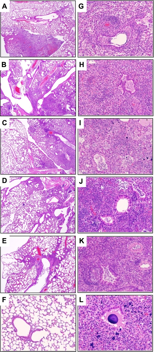
Fig 5.
Histopathologic changes in lungs of mice with secondary bacterial pneumonia. The images shown are H&E-stained sections of lungs collected at 7 dpi from BALB/c mice infected with 0.5 MLD50 of PR8 (A), WT H2N2 (B), ΔNA mutant H2N2 (C), WT H9N2 (D), or ΔNA mutant H9N2 (E) virus. All viruses carry the PR8 isogenic background. Original magnification, ×40. (F to M) Mice were infected with the indicated viruses and then challenged at 7 dpi with 104 CFU of pneumococci. Lungs were collected at 2 dppc. Representative H&E-stained sections are shown at ×200 magnification. (F) Mock-infected mice challenged with pneumococci. (G) Mice infected with PR8 and then mock challenged with PBS. The virus-S. pneumoniae groups correspond to WT H2N2 (H), ΔNA mutant H2N2 (I), WT H9N2 (J), ΔNA mutant H9N2 (K), and PR8 (L).

