Fig 3.
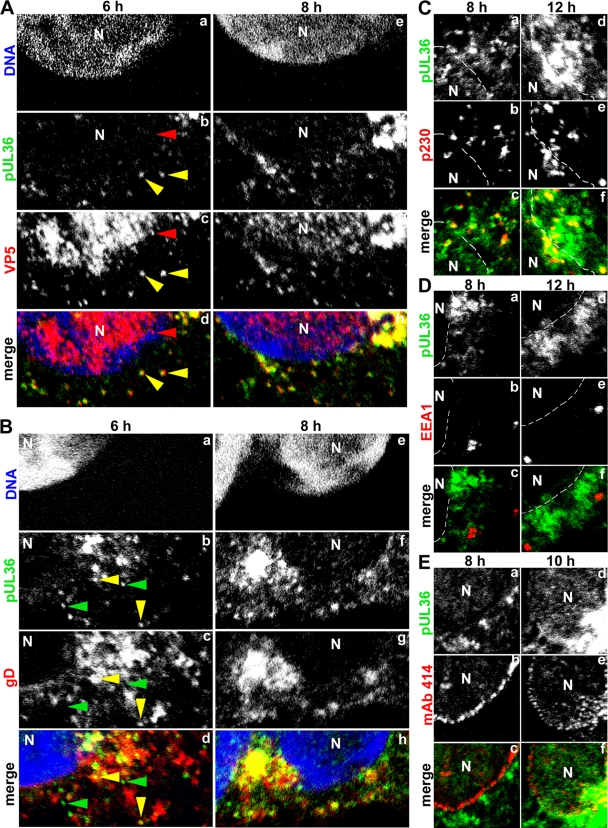
HSV1 pUL36 binds to progeny, cytosolic capsids prior to secondary envelopment. (A) Vero cells were infected with HSV1(17+) at an MOI of 10 PFU/cell and fixed with 3% paraformaldehyde at the indicated time points, followed by TX-100 permeabilization. The specimens were labeled with antibodies directed against pUL36 aa 1408 to 2112 (green; middle, pAb 147), the major capsid protein VP5 (A; red; MAb 5C10), the viral membrane protein gD (B; red; MAb DL-6), a marker for the trans-Golgi network (C; red; MAb α-p230), a marker for early endosomes (D; red; MAb α-EEA1), or the nuclear pores (E; red; MAb 414) and analyzed by confocal fluorescence microscopy. The nuclei (N) were labeled with the DNA stain TO-PRO-3 (blue in panels A and B) and are indicated by dashed lines (C to E).

