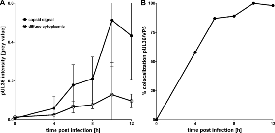
Fig 4.
Colocalization of pUL36 with cytoplasmic capsids. Vero cells infected with HSV1(17+) at an MOI of 10 PFU/cell were fixed at 4, 6, 8, 10, or 12 h postinfection and prepared for immunofluorescence using antibodies directed against pUL36 aa 1408 to 2112 (middle, pAb 147) and the major capsid protein VP5 (MAb 5C10) and TO-PRO-3, as for Fig. 3A. (A) For each time point postinfection, the pUL36 fluorescence intensity (gray value) was measured on cytoplasmic capsids (capsid signal) and the diffuse cytoplasmic signal (diffuse cytoplasmic) was determined. The error bars indicate the standard deviation. (B) These data were used to calculate the percentage of cytoplasmic capsids positive for pUL36.

