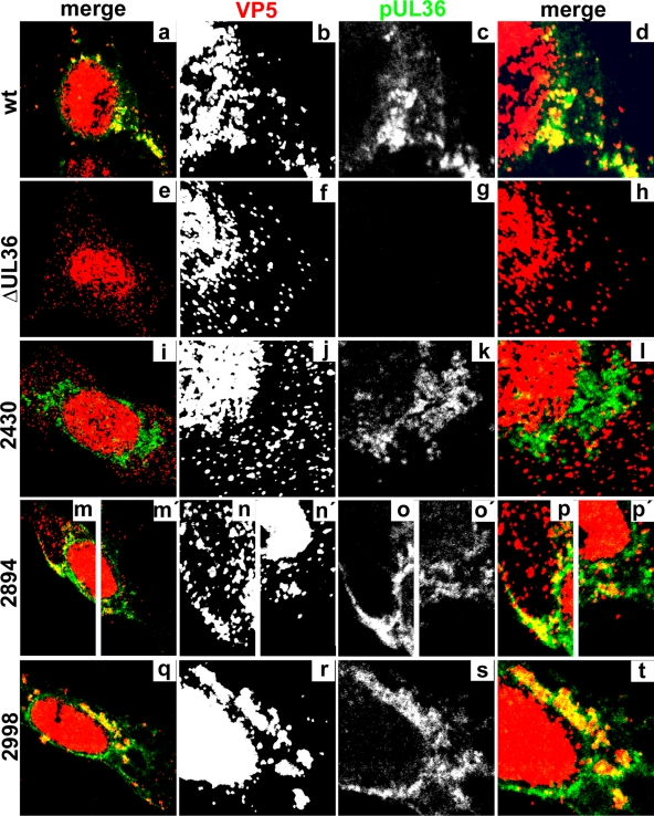
Fig 8.
Subcellular localization of truncated HSV1 pUL36 proteins. Vero cells were infected at an MOI of 0.2 PFU/cell with pHSV1(17+)blueLox (wt; a to d), with the mutant HSV1(17+)blueLox-ΔUL36 (ΔUL36; e to h), or with HSV1(17+)blueLox-UL36codon2430stop (2430; i to l), -UL36codon2894stop (2894; m to p and m′ to p′), or -UL36codon2998stop (2998; q to t), transcomplemented with pUL36 by amplification on Vero-HS30 cells, and fixed at 15 h p.i. with 3% paraformaldehyde, followed by TX-100 permeabilization. The specimens were labeled with antibodies directed against VP5 (red and b, f, j, n, n′, and r; MAb 5C10) or pUL36 aa 1408 to 2112 (green and c, g, k, o, o′, and s; middle, pAb 147) and analyzed by confocal fluorescence microscopy. Parts of the overviews (a, e, i, m, m′, and q) are shown at higher magnification in the horizontally neighboring panels.

