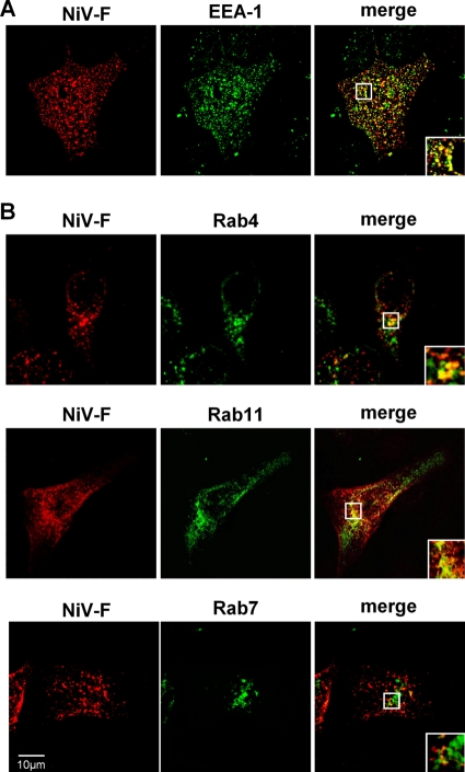
Fig 7.
Intracellular localization of internalized NiV F proteins in MDCK cells. (A) At 24 h p.t., an antibody uptake assay with NiV F-expressing MDCK cells was performed. Cells were incubated with a polyclonal anti-NiV serum for 1 h at 4°C and then incubated for 10 min at 37°C. Following endocytosis, surface-bound primary antibodies were blocked by incubation with a peroxidase-conjugated secondary antibody. After fixation and permeabilization, internalized F proteins were detected with AF 568-conjugated secondary antibodies. Early endosomes were stained with an anti-EEA-1 antibody and AF 488-conjugated secondary antibodies. (B) MDCK cells were cotransfected with pczCFG5-NiV-F and Rab4-CFP-, Rab11-CFP-, or Rab7-GFP-encoding plasmids. At 18 h p.t., surface-expressed F proteins were labeled with the anti-NiV serum. Then, endocytosis was allowed to proceed for 30 min at 37°C. Internalized F proteins were detected as described above. Images were recorded with a confocal laser scanning microscope (SP5; Leica). Insets represent expansions of the boxed regions of the merged images.

