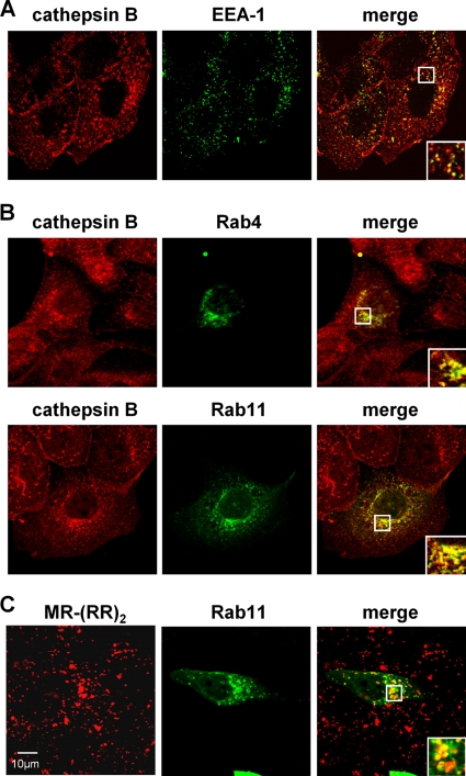
Fig 9.
Subcellular localization of endogenous cathepsin B in MDCK cells. (A) MDCK cells were fixed and permeabilized. Early endosomes were visualized with an EEA-1-specific primary antibody and AF 488-conjugated secondary antibodies. Endogenous cathepsin B was detected with anti-cathepsin B antibodies and AF 568-conjugated secondary antibodies. (B) MDCK cells were transfected with Rab4-CFP- or Rab11-CFP-encoding plasmids. At 18 h p.t., cells were fixed and endogenous cathepsin B was stained as described above. Images were recorded with a confocal laser scanning microscope (SP5; Leica). Insets represent expansions of the boxed regions of the merged images. (C) To visualize catalytically active cathepsin B, Rab11-CFP-expressing cells were incubated with the fluorogenic cathepsin B substrate Magic Red MR-(RR)2. Live cells were directly examined by confocal microscopy.

