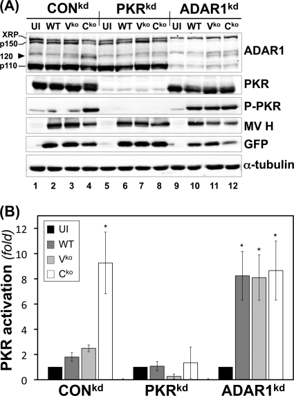
Fig 1.
ADAR1 deficiency enhances PKR activation following infection with WT, Vko, or Cko MV. (A) CONkd, PKRkd, and ADAR1kd cells were either left uninfected (UI) or infected with WT, Vko, or Cko MV as indicated. At 24 h postinfection, whole-cell extracts were prepared and analyzed by Western immunoblot assay with antibodies against ADAR1, PKR, phospho-Thr446-PKR, MV H, GFP, and α-tubulin. XRP marks the position of ADAR1 antibody-cross-reacting protein (mobility slower than that of p150); the arrowhead marks the postulated caspase-mediated p120 ADAR1 cleavage product. (B) Quantification of n-fold activation of PKR, as measured by the level of phospho-Thr446-PKR to total PKR, determined by Western immunoblot analysis as shown in panel A. *, P < 0.05 by Student t test for comparison of PKR activation in uninfected cells and cells infected with WT, Vko, or Cko MV. The results shown are the means and standard errors of three independent experiments.

