Fig 3.
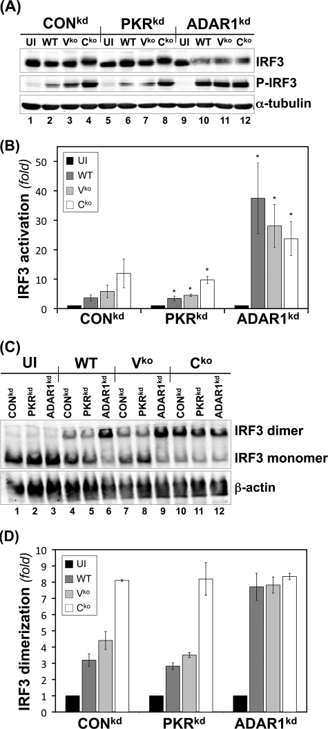
IRF3 activation is enhanced in ADAR1-deficient cells following infection with WT, Vko, or Cko MV. CONkd, PKRkd, and ADAR1kd cells were either left uninfected (UI) or infected with WT, Vko, or Cko MV as indicated. (A) At 24 h postinfection, whole-cell extracts were prepared and analyzed by Western immunoblot assay with antibodies against IRF3, phospho-Ser396-IRF3, and α-tubulin. (B) Quantification by Western immunoblot analysis of the n-fold activation of IRF3 expressed as the ratio of phospho-Ser396-IRF3 to total IRF3. *, P < 0.05 by Student t test for comparison of IRF3 activation in uninfected cells versus cells infected with the WT, Vko, or Cko virus. The results shown are means and standard errors (n = 3). (C) IRF3 activation as measured by dimer formation. Extracts were prepared and analyzed for IRF3 dimer formation by native PAGE as described in Materials and Methods. The blot was probed with antibody against IRF3 to detect dimer complexes fractionated from monomer protein. (D) Quantification of dimerization expressed as the ratio of IRF3 dimer to total IRF3 (monomer plus dimer). The results shown are means and standard deviations (n = 3).

