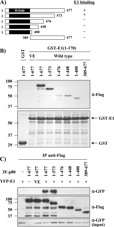
Fig 11.
E1 interacts with amino acids 1 to 573 of p80. (A) Schematic representation of the p80 truncations used for mapping the E1 binding domain on p80. The WD40 repeats of p80 are represented by black boxes. (B) In vitro pulldown assays. 3F-p80 truncations were expressed in rabbit reticulocyte lysates and tested for binding to the N-terminal domain of E1 (aa 1 to 170) fused to GST. GST alone or GST-E1 VE was used as a negative control. Bound proteins were detected by Western blotting using an anti-Flag antibody. At the bottom is shown a Coomassie blue-stained SDS-PAGE gel of the purified GST fusion proteins used in these pulldown experiments. The sizes of molecular weight markers (in thousands) are indicated on the left. (C) In vivo coimmunoprecipitation assays. The indicated 3F-p80 truncations were expressed in C33A cells together with YFP-E1 and immunoprecipitated using anti-Flag antibodies. The presence of YFP-E1 in the immunoprecipitates was determined by Western blotting using an anti-GFP antibody. Cells expressing the YFP-E1 VE mutant protein and full-length 3F-p80 were used as a negative control.

