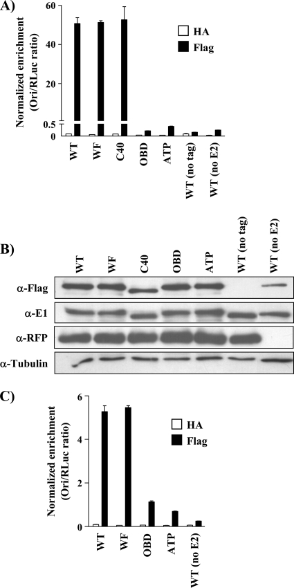
Fig 3.
The p80-binding domain is not required for the stable assembly of E1 at the viral origin. (A) ChIP assays were performed in C33A cells transfected with 3F-E1 (WT or mutant), RFP-E2, and an origin plasmid (pFLORI31). Equal quantities of cell lysates were immunoprecipitated with anti-Flag (E1) or anti-HA (nonspecific) antibodies. The results of the ori enrichment levels determined by qPCR are shown after normalization to input DNA, using the internal control pRL (RLuc). Each value is the average of at least two replicates, with the standard deviations presented as error bars. (B) Western blot showing the expression of E1 mutant proteins detected by either the anti-Flag or anti-E1 antibody and of RFP-E2 using an anti-RFP antibody. β-Tubulin was used as a loading control. (C) C33A cells were transfected with plasmids expressing 3F-E1 (WT or mutant) and RFP-E2 and one containing the origin (pFLORI31). The cells were treated with 5 μg/ml of aphidicolin 4 h posttransfection, and ChIP assays were performed on the lysates using anti-Flag and anti-HA antibodies. The results of the ori enrichment levels determined by qPCR are shown after normalization to input DNA using the internal control pRL (RLuc). Each value is the average of at least two replicates, with the standard deviations presented as error bars.

