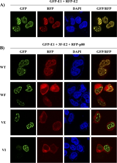
Fig 4.
p80 is relocalized to nuclear foci in an E1- and E2-dependent manner. (A) Fluorescence confocal microscopy images showing the subcellular localization of GFP-E1 and RFP-E2. (B) Subcellular localization of RFP-p80 when coexpressed with 3F-E2 and WT or p80-binding-defective GFP-E1. Nuclei were stained with DAPI.

