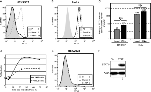
Fig 4.
The BST2 promoter is activated by type I IFN with a delayed kinetics. HEK293T (A) and HeLa (B) cells were treated with IFN-α for 24 h, and BST-2 cell surface expression was analyzed by flow cytometry. (C and D) Activity of the BST2 promoter relative to the β-actin promoter. pGL4.17-BST2_ffLuc(FL) and pGL4.70-Actin_renLuc (200 ng each) were cotransfected in duplicate in either HEK293T or HeLa cells (105 cells). (C) Samples were collected under basal conditions or after 24 h of IFN-α treatment. The luciferase signal obtained with pGL4.70-Actin_renLuc was set as 1.0 (numbers above the bars represent fold increase of promoter activation by IFN-α). (D) Samples were collected at different time intervals post-IFN-α treatment and analyzed for dual-luciferase activity. Firefly luciferase activity driven by the BST2 promoter was expressed relative to control Renilla luciferase activity. Fold increase was calculated relative to the value obtained at time zero, which was set at 1. (E) HEK293T cells transfected with a STAT1-encoding plasmid (1 μg of plasmid per 2 × 10⁵ cells) were analyzed for BST-2 cell surface expression by flow cytometry, 48 posttransfection. (F) Western blot of whole-cell extracts from the indicated transfected cells revealed with α-STAT1 and α-actin Abs. The data shown are representative of results obtained in at least three independent experiments.

