Fig 2.
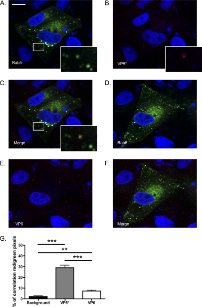
(A to C) UK VP5* associated with the early endosome marker Rab5-EGFP in MA104 cells at 15 min postinfection. Colocalization events are highlighted by white boxes that are shown at higher magnification at the bottom right of the pictures. (D to F and G) UK VP6 was significantly less associated with Rab5 than VP5*. Bar, 10 μm. (G) Average percentage of red pixels (UK+) that are also green (Rab5+) under each condition for each field. ***, P < 0.001; **, P < 0.01 (one-way ANOVA test followed by the Newman-Keuls multiple-comparison posttest).

