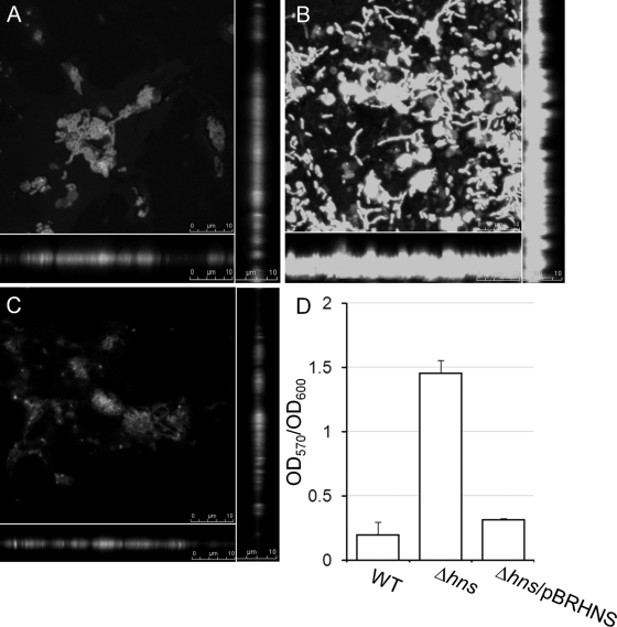
Fig 1.
Repression of biofilm formation by H-NS. Strains C7258ΔlacZ (A), AJB80 (Δhns) (B), and AJB80 complemented with plasmid pBRHNS (C) were allowed to form static biofilm in LB medium, stained with Syto-9, and examined by confocal microscopy. The center of each panel is an XY section through the biofilm. Vertical sections through the biofilm are shown to the right and bottom. (D) Quantification of biofilm formation using the crystal violet stain.

