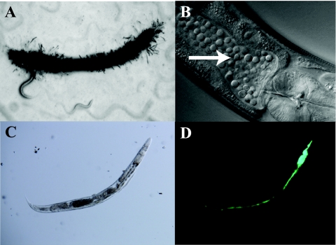
Fig 1.
Infections of C. elegans. Microscopic images of various infections of C. elegans. are shown. (A) D. coniospora infection in a wild-type worm at day 2. Note the characteristic hyphal penetration throughout the animal. (B) C. neoformans infection in a wild-type worm at day 5, where the intestine is packed with proliferating yeast cells (arrow). (C and D) Bright-field (C) and fluorescence (D) images of a representative wild-type animal at day 3 of an S. Typhimurium infection (with green fluorescent protein used for detection). Here, the pharyngeal structure has been destroyed by the infection and the intestine is distended and full of bacteria. (Panels C and D were reprinted from reference 44.)

