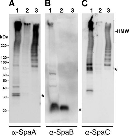
Fig 1.
Western analysis of fractionated GG cells and affinity-purified pili. Membranes were probed with polyclonal rabbit antisera against recombinant SpaA (A), SpaB (B), or SpaC (C). The wells were loaded with cell wall-associated extract (lane 1), cell-free extract (lane 2), or affinity-purified pili (lane 3). The calculated position of the corresponding monomeric pilin is indicated with an asterisk on the right side of each panel.

