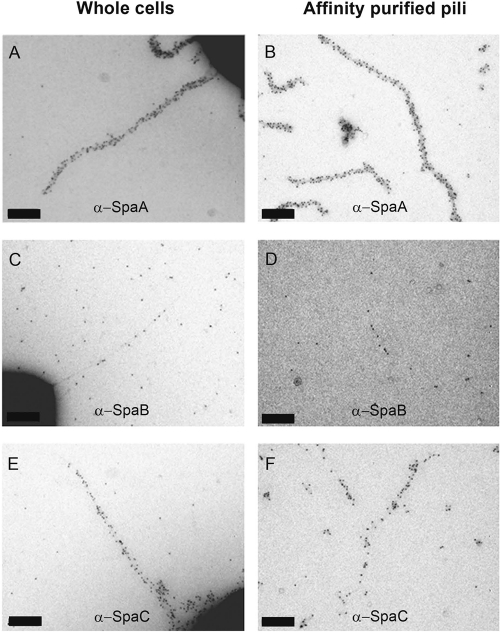
Fig 3.
TEM images of single-labeled GG cells and affinity-purified SpaCBA pili. Representative images of single-labeled cell wall-associated pili (A, C, and E) or affinity-purified pili (B, D, and F). The pili were labeled with SpaA (A and B), SpaB (C and D), or SpaC antiserum (E and F), followed by the addition of 5-nm pAg. Scale bars indicate 100 nm.

