
Fig 5.
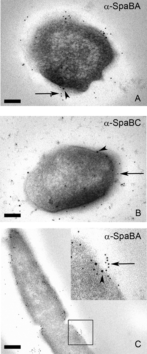
TEM images of immunogold-labeled GG thin sections. Whole GG cells first were labeled with SpaB antibodies followed by 10-nm protein A gold particles (arrowheads). The pili then were labeled with SpaA (A and C) or SpaC antiserum (B) and 5-nm pAg (arrow). The area enclosed in the black square in panel C is highlighted in the upper right corner of the panel. Scale bars, 100 (A and B) or 200 nm (C).