
Figure 1.
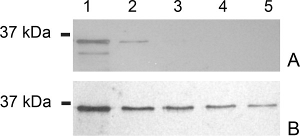
Western blot detection of Sapp1p associated with the cell surface of C. parapsilosis. Panel A: fractions obtained by cell surface washing with PTB buffer; Panel B: fractions from washing with PTB containing 0.5% βME. Lane 1, fraction after 10-min incubation; Lanes 2–5, fractions after repeated washings. Detection was performed using polyclonal antibodies raised against Sapp1p.