
Figure 1.
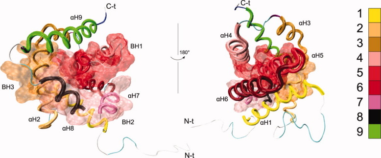
Bax 3D structure (PDB ID: 1F16). The nine α-helices are shown as tubes. The location of BH1-3 is indicated by the transparent surfaces. Labels indicate the N-terminal (N-t) and C-terminal (C-t) ends of the protein. [Color figure can be viewed in the online issue, which is available at wileyonlinelibrary.com.]